Overview
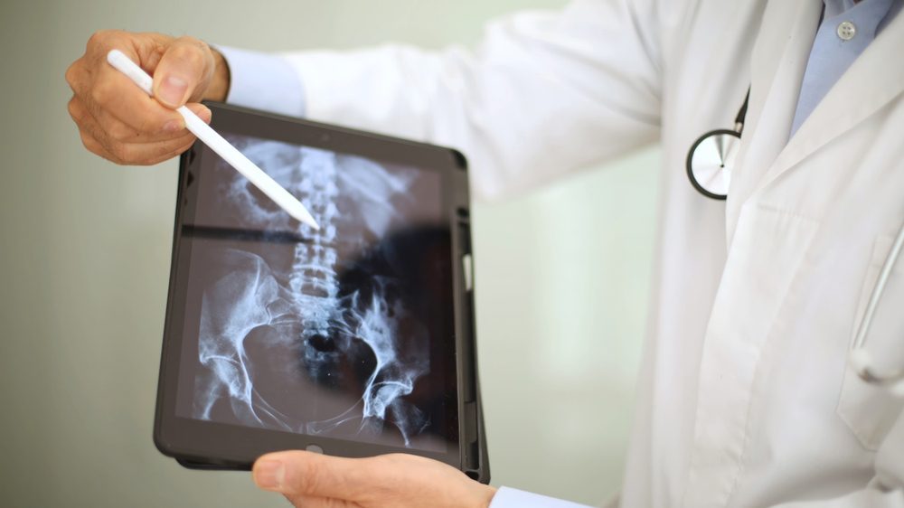
Radiological assessments are vital diagnostic tools that utilize various imaging techniques to visualize the internal structures of the body. These assessments help healthcare providers diagnose medical conditions, monitor treatment progress, and plan surgical interventions. Radiological imaging includes X-rays, computed tomography (CT) scans, magnetic resonance imaging (MRI), ultrasound, and nuclear medicine, each offering unique insights into a patient’s health status.
Indications for Radiological Assessments
Radiological assessments are indicated for a variety of clinical scenarios, including:
- Diagnostic Imaging: Identifying fractures, tumors, infections, or other abnormalities within the body.
- Preoperative Planning: Assessing anatomical structures before surgical procedures.
- Monitoring Disease Progression: Evaluating changes in the size or appearance of lesions or tumors over time.
- Guiding Therapeutic Procedures: Assisting in interventions such as biopsies or catheter placements.
- Assessing Treatment Response: Evaluating the effectiveness of therapies, such as chemotherapy or radiation.
Types of Radiological Assessments
- X-rays:
- Standard X-rays: Commonly used to visualize bones and detect fractures, dislocations, or degenerative conditions.
- Fluoroscopy: Provides real-time imaging of moving body parts, often used in gastrointestinal studies.
- Computed Tomography (CT) Scans:
- CT Scans: Provide cross-sectional images of the body, useful for detailed examination of complex structures, such as the brain, abdomen, and chest.
- CT Angiography: Visualizes blood vessels to assess for blockages or abnormalities.
- Magnetic Resonance Imaging (MRI):
- MRI Scans: Utilize powerful magnets and radio waves to create detailed images of soft tissues, making them ideal for evaluating the brain, spinal cord, muscles, and joints.
- Functional MRI (fMRI): Measures brain activity by detecting changes in blood flow, often used in neurological studies.
- Ultrasound:
- Diagnostic Ultrasound: Uses sound waves to create images of soft tissues, often employed in obstetrics, gynecology, and musculoskeletal evaluations.
- Doppler Ultrasound: Assesses blood flow in vessels and can detect abnormalities in blood circulation.
- Nuclear Medicine:
- PET Scans (Positron Emission Tomography): Assess metabolic activity and detect cancer by using radioactive tracers.
- SPECT Scans (Single Photon Emission Computed Tomography): Provide functional information about organs, useful in cardiac and neurological evaluations.
Radiological Assessment Procedure
- Pre-Assessment Preparation:
- Patients may receive specific instructions regarding food and fluid intake, medication use, or clothing choices before the imaging procedure.
- Imaging Procedure:
- Patients are positioned appropriately for the imaging study, and technologists operate the imaging equipment while ensuring patient comfort and safety.
- Imaging may involve multiple views or angles to capture comprehensive data.
- Radiologist Interpretation:
- After the imaging study, a radiologist analyzes the images and prepares a report detailing findings, conclusions, and recommendations.
- Results Communication:
- The radiologist’s report is shared with the referring physician, who discusses the findings with the patient and formulates a management plan based on the results.
Benefits of Radiological Assessments
- Accurate Diagnosis: Radiological imaging provides essential information for accurate diagnosis and treatment planning.
- Non-Invasive: Most imaging techniques are non-invasive and allow for detailed internal visualization without the need for surgery.
- Comprehensive Evaluation: Radiological assessments can examine multiple body systems and structures simultaneously, providing a holistic view of a patient’s health.
- Real-Time Guidance: Some imaging modalities, such as ultrasound and fluoroscopy, provide real-time feedback during procedures, enhancing safety and effectiveness.
Possible Risks and Limitations
- Radiation Exposure: Some imaging techniques, such as X-rays and CT scans, involve exposure to ionizing radiation, though the benefits typically outweigh the risks.
- Contrast Reactions: Use of contrast agents in some imaging studies may carry a risk of allergic reactions or kidney issues in certain patients.
- Access and Availability: Availability of advanced imaging technologies may vary by facility, potentially impacting timely diagnosis.
Final Results
With the implementation of radiological assessments, patients can expect:
- Informed Clinical Decisions: Radiological findings provide critical insights that help guide diagnosis and treatment strategies.
- Timely Interventions: Quick access to imaging results can lead to timely interventions and improved patient outcomes.
- Enhanced Monitoring: Ongoing radiological evaluations allow for continuous monitoring of health conditions, facilitating proactive management.

